Figure 12 3 Floor Of The Cranium

The Base Of The Skull Superior View Of Cranial Floor Anatomy Images Illustrations Anatomy Images Charac Craniosacral Therapy Anatomy Anatomy Coloring Book

This Figure Shows The Structure Of The Cranial Fossae The Top Panel Shows The Superior View Anatomy And Physiology Textbook Human Skull Anatomy Anatomy Bones

Right Orbit Bones Human Anatomy And Physiology Rectus Muscle Head And Neck

Foramen Magnum Hypoglossal Canal Jugular Foramen Anatomy Superior View Of The Skull Skull Anatomy Anatomy Bones Medical Anatomy

Pin On Daily Cao

May 2009 Skull Anatomy Medical Anatomy Anatomy

A rapid review of the internal structures of the cranium and the exit points of the 12 cranial nerves.
Figure 12 3 floor of the cranium.

Part 1 The Axial Skeleton 71 The Skull Consists Of 8 Cranial Bones And 14 Facial Bones Human Anatomy And Physiology Dental Anatomy Medical Anatomy Anatomy Bones
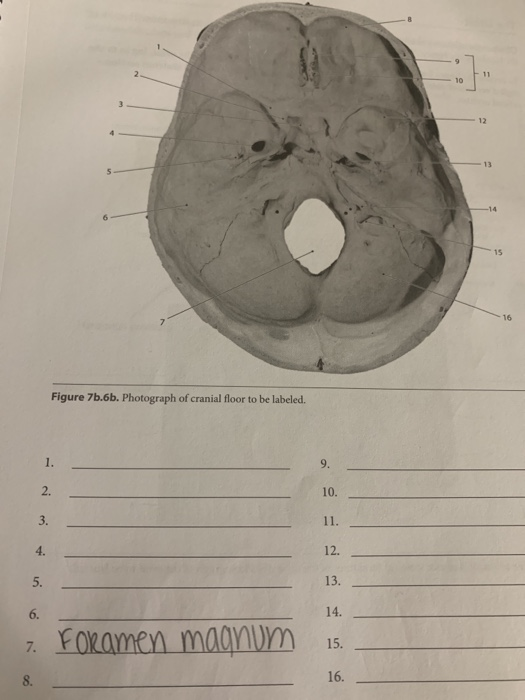
Solved 11 10 3 12 13 14 15 16 Figure 7b 6b Photograph Of Chegg Com

Pin By C Puckett Animal Lives Matter On Cn Vii Facial Human Brain Anatomy Cranial Nerves Brain Anatomy

Skull Inferior View With The Mandible Removed Skull Anatomy Anatomy And Physiology Palatine Bone
Source : pinterest.com
